Most otoplasty patients heal well and love their results. But in a small percentage of cases, the ears don’t heal as planned — the fold may loosen over time, asymmetry can become noticeable, or too much cartilage was removed, leaving an unnatural contour. When the outcome falls short of what you hoped for, otoplasty revision is used to correct it. Revision surgery is more delicate than the first procedure because the cartilage has already been reshaped and scar tissue has formed, so it requires a surgeon with specific experience in secondary ear corrections. Timing matters too: most surgeons recommend waiting at least six months after the initial surgery before considering a revision, since early swelling can mask the true result. Knowing when a revision is genuinely needed and when patience is better is the first step toward a satisfactory result.
What Is Otoplasty Revision and How Does It Differ From the Initial Surgery?
Otoplasty itself is a relatively straightforward procedure that reshapes the cartilage of the ear, usually to reduce prominence or correct asymmetry. When the first result falls short — whether due to healing quirks, technique, or tissue behavior — a revision otoplasty becomes the path forward. It is corrective surgery performed on an ear that has already been operated on, with the goal of refining, repairing, or completely redoing the original work.
The difference between the two procedures goes far deeper than simply “operating again.” During the initial surgery, the surgeon works with untouched anatomy: predictable cartilage, clear tissue planes, and a standard surgical roadmap. In a revision, that same ear presents scar tissue, altered blood supply, stiffened or weakened cartilage, and sometimes cartilage that was over-resected the first time. These changes demand a different mindset and often a different technique.
Revision work also tends to be more individualized. The surgeon must first analyze what went wrong — an overcorrection, an undercorrection, an exaggerated antihelical fold, a visible scar, or a relapsed protrusion — and then decide whether to reconstruct using remaining cartilage, borrow graft material from the rib or the ear itself, or release existing scarring to reposition the ear naturally. Because of this added complexity, revision procedures frequently take longer in the operating room and require a surgeon with specific experience in secondary ear surgery.
For patients, understanding this distinction matters: a revision is not a repeat of the first operation, but a more technically demanding procedure tailored to anatomy that has already been altered once.
Common Reasons Patients Seek Ear Surgery Correction
When an initial ear pinning procedure doesn’t deliver the outcome a patient hoped for, the disappointment is often paired with confusion: what exactly went wrong? In practice, the reasons people pursue revision otoplasty tend to fall into a handful of recognizable patterns, most of which stem from technique, healing biology, or the natural behavior of cartilage over time.
The most frequently seen issues include:
- Residual or recurring prominence — the ear drifts back toward its original position, often because cartilage memory overpowered the correction or sutures loosened during healing.
- Asymmetry between the two ears — one ear sits noticeably higher, flatter, or closer to the head than the other, a discrepancy that becomes more bothersome once the swelling settles.
- Overcorrection — the ear is pinned too aggressively, sitting unnaturally flat against the head, sometimes described as a “glued-down” appearance.
- Visible or hypertrophic scarring — thickened, raised, or darkened scars behind the ear that draw attention rather than hiding discreetly in the natural crease.
- Contour irregularities — sharp edges, buckling of the antihelical fold, or a “telephone ear” deformity where the upper and lower poles protrude while the middle is over-tightened.
It’s worth understanding that some of these outcomes aren’t necessarily surgical errors. Cartilage is a living tissue with elasticity and memory, and even a technically excellent operation can be reshaped by the healing process. Younger patients, whose cartilage is softer and more springy, may experience more drift than adults. Similarly, minor asymmetry is present in nearly all natural ears, so the goal of any correction is improvement within a realistic range — not flawless mirror-image perfection.
There’s also a subset of patients seeking correction years after their first surgery, when results that once looked good gradually changed. This is why identifying the *cause* of the problem matters: the reason behind the dissatisfaction directly shapes which corrective approach a surgeon will recommend.
Signs That Your Ear Results May Need a Revision
Recognizing when your ears haven’t healed the way you hoped is the first step toward deciding whether a follow-up procedure makes sense. While some swelling and subtle asymmetry are completely normal during the early months, certain findings tend to persist and signal that a revision otoplasty may be genuinely warranted rather than simply premature concern.
One of the clearest indicators is when one or both ears gradually return toward their original position months after surgery. This “ear springing back” usually means cartilage memory overcame the correction, and it rarely improves on its own. Similarly, if the gap between your ears and the sides of your head remains noticeably uneven once healing has stabilized, that persistent asymmetry is unlikely to resolve without further intervention.
Contour issues deserve attention too. Sharp edges, unnatural folds, or a “pinned back” appearance that looks overcorrected can affect how you feel about your results just as much as residual prominence. Visible scarring behind the ear — especially thick, raised, or discolored scars — is another legitimate reason to seek evaluation, since mature scars that remain prominent after a full year typically won’t fade further.
Pay attention to functional complaints as well. Tenderness when sleeping, sensitivity to cold, or discomfort when wearing glasses can point to structural problems that surgery may address.
Finally, trust how the results look in photographs. Mirror checks can be misleading due to angle and lighting, but comparing consistent photos over time gives you a more honest picture of whether the improvement you expected has actually lasted.
How Long Should You Wait Before Considering a Second Procedure?
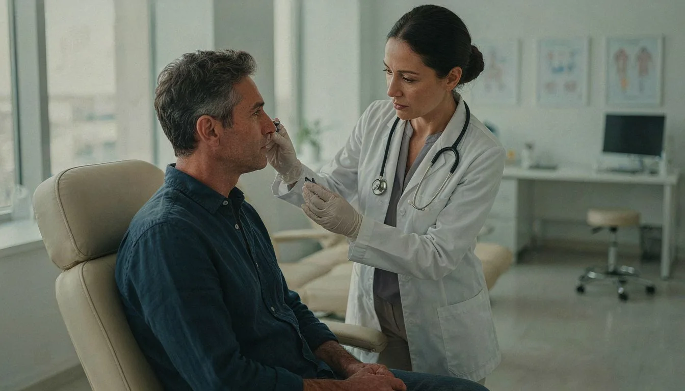
Timing is one of the most important decisions in the entire revision process. Surgeons generally recommend waiting at least six months after your initial otoplasty before evaluating whether a second procedure is truly necessary, and many prefer a full twelve months for complex cases. This waiting period exists for a practical reason: ears change significantly as they heal.
During the first few months, swelling, firmness, and slight asymmetry are completely normal. Cartilage, in particular, softens and settles gradually, and what looks like residual or recurring prominence at three months may improve on its own by month six or seven. Scars also mature over this period, often fading and flattening considerably without any intervention.
Patience protects you in two ways. First, it prevents unnecessary surgery on tissue that is still remodeling. Second, it allows your surgeon to operate on ears that have reached a stable state, which makes the revision more predictable and technically easier. Operating on inflamed or swollen cartilage increases the risk of complications and can compromise the final result.
Waiting has limits. If you are experiencing pain, infection, skin loss, or a suture working its way through the skin, these issues should be addressed promptly rather than delayed until the six-month mark. Your surgeon will distinguish between problems that threaten healing and purely cosmetic concerns that benefit from time.
One exception worth knowing: some surgeons perform a very early “touch-up” of minor asymmetries within the first few weeks, while the tissue planes are still soft. This is a selective approach, not the standard route, so discuss the timing strategy openly during your evaluation.
What Happens During the Revision Consultation and Evaluation
The consultation is arguably the most decisive step in any revision journey, because unlike a first-time procedure, the surgeon is working with tissue that has already been operated on. Scar tissue, altered cartilage memory, and changes in blood supply all influence what is surgically possible. That is why a thorough evaluation goes far beyond a quick glance at the ears — it is a structured assessment designed to find out what went wrong, why it happened, and what can realistically be achieved the second time around.
A strong revision consultation typically includes several core components:
- Detailed medical and surgical history: The surgeon reviews your original operative report if available, the technique that was used, and how your healing progressed. Knowing whether cartilage was cut, sutured, or scraped helps predict how it will behave now.
- Physical examination of both ears: Cartilage firmness, skin thickness, scar quality, and blood supply are assessed by touch as well as visually. Palpation often reveals problems like suture knots or cartilage edges that photos cannot show.
- Standardized photographs: Front, side, and angled images document your current asymmetries and contours, serving as both a planning tool and a baseline for measuring improvement.
- A conversation about your goals: You will be asked what specifically bothers you — whether it is the degree of correction, symmetry, scarring, or contour — so expectations can be aligned with surgical reality.
- Timing assessment: The surgeon confirms whether enough healing time has passed since your original otoplasty, since revising immature tissue compromises both safety and results.
Beyond these basics, expect an honest discussion of risk. Revisions carry a somewhat higher chance of complications than primary otoplasty, and a trustworthy surgeon will explain this plainly rather than minimize it. Some may also use digital imaging or computer simulation to illustrate possible outcomes, though you should treat these as directional guides rather than guarantees.
Finally, you should leave the consultation with a clear written plan: the proposed technique, the estimated revision cost, anesthesia details, and the recovery timeline. If a surgeon cannot articulate why your first result failed and how their approach differs, consider seeking a second opinion before committing to another operation.
Surgical and Non-Surgical Options for Correcting Ear Shape
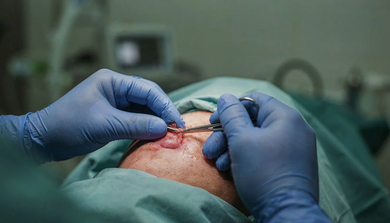
Once your evaluation is complete, the next question is how the correction will actually be achieved. The good news is that a disappointing ear result rarely means you’re out of options — it simply means choosing the right technique for the specific problem.
Most structural issues call for a surgical approach. When cartilage has springs back, sutures have failed, or excess cartilage creates sharp edges, your surgeon may recommend a revision procedure that repositions, trims, or reinforces the underlying framework. Techniques vary: some revisions use fresh internal sutures, others rely on cartilage grafting harvested from the ear itself or the rib, and severe cases may benefit from reshaping the ear’s fold entirely. Because tissue from the first operation is often scarred and less pliable, a surgical revision typically demands greater precision and experience than the original otoplasty — which is why choosing a surgeon who regularly performs secondary ear surgery matters so much.
Not every concern requires returning to the operating room. Minor asymmetries, small contour irregularities, or subtle scar concerns can sometimes be managed without incisions. Injectable treatments such as corticosteroids can soften raised or hypertrophic scars, and in carefully selected cases, tiny amounts of filler can camouflage mild surface irregularities. There are also minimally invasive options like thread-based repositioning, though these are appropriate only for specific anatomy and tend to offer less predictable long-term stability than surgery.
Your surgeon will weigh several factors when recommending a path: how much healing time has passed, the quality of the remaining cartilage, the amount of skin available, and of course what you’re hoping to achieve. A patient bothered primarily by a small scar will have a very different plan than one whose ear has partially unfolded again.
It’s worth knowing that revision surgery often takes less time than the initial procedure — many are completed in one to two hours under local or light anesthesia — but the refined techniques involved make careful planning far more important than speed.
Recovery Expectations and Long-Term Results After Revision Otoplasty
Recovery after a second ear surgery tends to be gentler than most patients expect, largely because revision otoplasty usually involves smaller, more targeted corrections than the original procedure. Healing tissue behaves differently the second time around, and knowing what lies ahead helps you pace yourself realistically.
In the first week, expect mild swelling and some bruising around the ears. A protective headband is typically worn continuously at first, then at night for several weeks, to keep the ears in their new position while the cartilage settles. Discomfort is usually modest and well managed with prescribed medication. Most patients return to desk work or school within seven to ten days.
Weeks two through six bring steady improvement. Swelling continues to subside, the ears soften in appearance, and strenuous activity is gradually reintroduced. Contact sports and anything that risks bending the ears remain off-limits for at least six to eight weeks, since newly shaped cartilage is vulnerable to pressure during this window.
One honest caveat deserves mention: because revision work often involves scar tissue and a limited blood supply, healing can take slightly longer than after the first operation. Small areas of firmness or subtle asymmetry may persist for several months before relaxing into their final form. Patience during this phase is genuinely part of the treatment.
The long-term picture is encouraging. When the underlying cause was correctly identified during the evaluation and the appropriate technique was chosen, results from an ear surgery correction are typically stable and permanent. Cartilage does not drift back over time once it has fully healed, so the vast majority of revision patients enjoy their refined ear shape for life. Satisfaction rates are consistently high, particularly among patients whose concerns were asymmetry or residual prominence rather than complex structural problems.
To protect your investment long-term, follow your surgeon’s instructions precisely, attend all follow-up appointments, and avoid sleeping directly on the ears during the early months. With realistic expectations and proper aftercare, the outcome of a carefully planned revision can finally deliver the balanced, natural-looking ears the first surgery missed.
FAQs
How soon after otoplasty can a revision be safely performed?
Most surgeons recommend waiting at least 6 to 12 months after the initial otoplasty before performing a revision. This waiting period allows the swelling to fully subside, the cartilage to settle into its new shape, and the tissues to heal completely, so the surgeon can accurately assess what actually needs correcting. Operating too early risks revising a problem that would have resolved on its own, or working with tissue that is still fragile. If the issue is minor, some surgeons may act sooner, but for most cases the full healing period gives the best and safest results.
What are the most common reasons for otoplasty revision after otoplasty?
The most common reasons for otoplasty revision are recurrence of ear protrusion, asymmetry between the two ears, and an overly corrected or "pinned back" appearance. Other frequent causes include visible or hypertrophic scarring, suture-related issues such as protruding threads, and residual cartilage irregularities. Many of these problems stem from insufficient correction during the initial surgery, technique limitations, or natural healing responses like cartilage springing back, which is why a skilled and experienced surgeon significantly lowers revision risk.
Is the recovery faster for a revision than for the initial surgery?
No, recovery from a revision otoplasty is often a bit different rather than strictly faster. Because the surgeon is working with scar tissue and a previously operated ear, swelling and tenderness can sometimes last slightly longer than after the first surgery. Many patients find the overall experience easier the second time, since they already know what to expect and the aftercare routine feels familiar. Most people are back to work and light activities within about a week, just as with the initial procedure.